
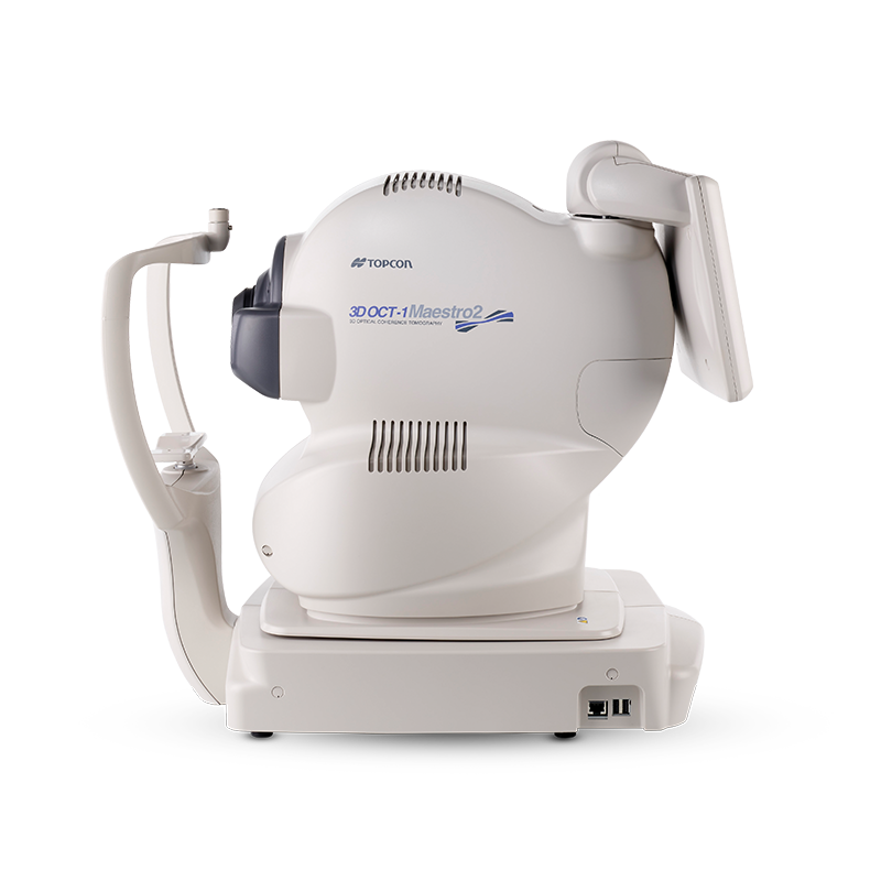
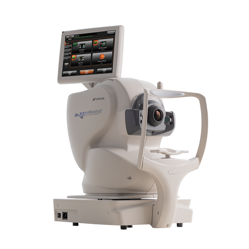
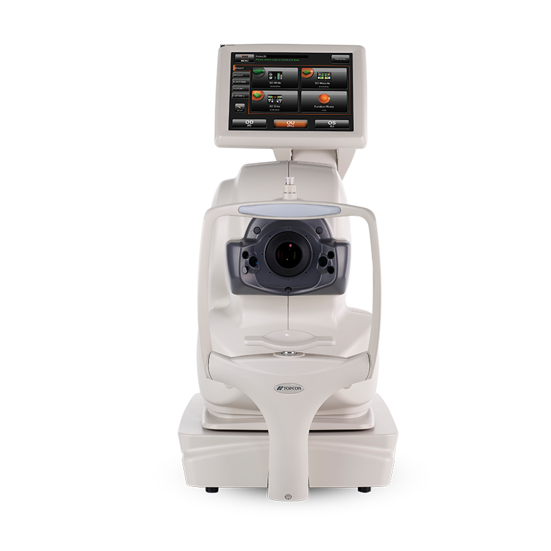
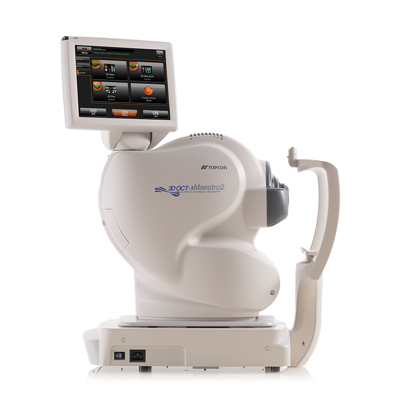
Topcon 3D OCT-1 Maestro2 Spectral Domain OCT
An Integrated Platform for High Quality Imaging and Efficient Data Management
With the Topcon Maestro2, you have fast, multi-modality OCT/Fundus imaging, and advanced data management. The Maestro2 is the complete clinical workstation for any busy practice. With a single touch, the Maestro2 automatically performs alignment, focus, optimizing and capturing. After capturing, the report can be immediately displayed by clicking on the icon. In addition to automated capture, the Maestro2 offers manual/semi-manual options for difficult-to-image patients.
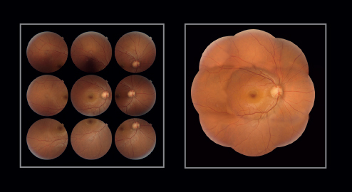
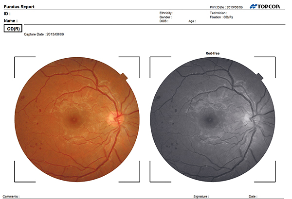
The Maestro2 has an integrated full-color fundus camera. With one touch, you can simultaneously acquire a posterior OCT image and a true color fundus image. This allows for PinPoint Registration and structural confirmation of the pathology. A small pupil function is also available, as well as fundus only capture.
BENEFITS:
Automation - With the touch of a button, Maestro enables you to acquire a high resolution OCT image and a true-color fundus photo. Auto Focus, Auto Alignment, Auto Capture
Speed of Use - Maestro requires nothing more than to touch the capture icon and Start Capture button. Alignment, focus, optimizing, and capturing are performed in an automatic procedure. After the capturing, a report can be immediately displayed by clicking on the icon.
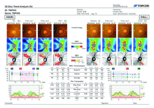
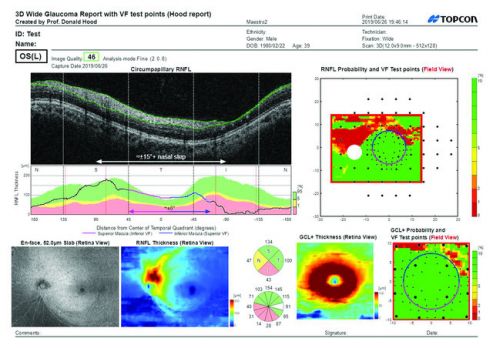
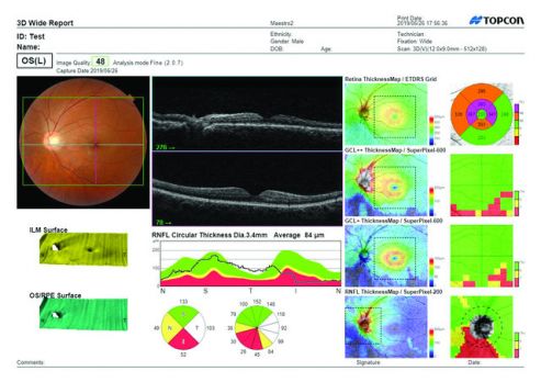
3D Wide12x9mm, The Hood Report for Glaucoma with Reference Database - Retinal Thickness/RNFL/GCL and Optic Nerve Metrics in just one scan. Additionally, the New Hood Report for Glaucoma is available; an innovative one-page report that simplifies your structure/function decision-making.
Connectivity - Connect your Topcon and other diagnostic instruments, whether they are DICOM or not to review your exam data on any PC or mobile device.
Fully-Featured OCT With True Color Fundus Imaging - Not Just an OCT: Powerful, Versatile and Fast (50K A-Scans/sec.) OCT Fundus Camera that captures beautiful and sharp B-Scans, wide-encompassing disc/macula 12x9 cubes, and informative anterior segment scans - all of which are simultaneously and easily obtained with a true color image.
Available:
- New
Combination OCT and true color fundus
Compact and space-saving design
The NEW Hood Report for Glaucoma
Reference database for retina, RNFL, GCL+, and GCL++ thickness
Automatic layer segmentation
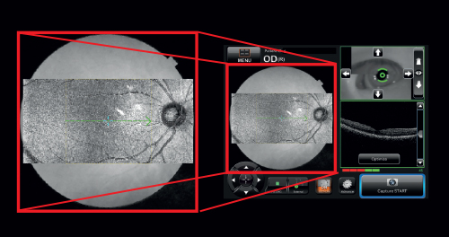
Widefield OCT
Anterior segment OCT
Panoramic fundus imaging
Observation & photographing of the fundus
Type of photography: Color, Red-free (Note 1) & IR (Note 3)
Picture angle for photography: 45° ± 5% or less, 30° or equivalent (digital zoom)
Operating distance: 34.8 ± 0.1mm (when taking a picture of fundus)
Photographable diameter of pupil:
Normal pupil diameter: ø4.0mm or more, Small pupil diameter: ø3.3mm or more
Fundus image resolution (on fundus):
Center : 60 lines/mm or more
Middle (r/2) : 40 lines/mm or more
Middle (r) : 25 lines/mm or more
IR photography : Center: 5 lines/mm or more (Note 3)
Observation & photographing of the fundus tomogram
Scan range (on fundus) Horizontal direction: 3 – 12mm ± 5% or less
Vertical direction: 3 – 9mm ± 5% or less
Scan pattern: 3D scan (horizontal/vertical), Linear scan (Line-scan/Cross-scan/Radial-scan)
Scan speed: 50,000 A-Scans per second
Lateral resolution: 20μm or less
In-depth resolution: 6μm or less
Photographable diameter of pupil: ø2.5mm or more
Observation & photographing of the fundus image/fundus tomogram
Fixation target:
Internal fixation target: Dot matrix type organic EL, The display position can be changed and adjusted. The displaying method can be changed.
Peripheral fixation target: This is displayed according to the internal fixation target displayed position.
External fixation target
Observation & photographing of anterior segment
Type of photography: Color & IR (Note 3)
Operating distance: 62.6 ± 0.1mm (when taking a picture of anterior segment) (Note 2)
Observation & photographing of the anterior segment tomogram
Operating distance: 62.6 ± 0.1mm (when taking a picture of anterior segment) (Note 2)
Scan range: (on cornea) (Note 2)
Horizontal direction: 3 – 6mm ± 5% or less
Vertical direction: 3 – 6mm ± 5% or less
Scan pattern: Linear scan (Line-scan/Radial-scan)
Scan speed: 50,000 A-Scans per second
Fixation target: External fixation target
(Note 1) Digital Red-free photography that processes a color image and displays it in pseudo-red-free condition.
(Note 2) Observation & photography of anterior segment can be performed only when the anterior segment attachment (HA-2) is used.
(Note 3) This is used only for recording the position where a tomogram is captured.
As a division of the Advancing Eyecare Alliance, Enhanced Medical Services is proud to provide financing options for all of your equipment needs.
Through Advancing Eyecare Finance, take advantage of multiple finance structures designed to meet your budget and ownership goals.
Request A Quote
Our team is dedicated to helping you find the right equipment to fit your needs and budget. Simply fill out the form below and one of our experienced staff will get back with you promptly with a quote. If you don't see what you're looking for, be sure to ask as new inventory is arriving daily.